The Brain Building Breakthrough
Axolotls, a species of salamanders native to lakes near Mexico City, have long captivated the scientific community due to their extraordinary regenerative abilities. These amphibians can regenerate entire limbs, spinal cord, heart and even parts of their brain. However, the molecular mechanisms that allow such remarkable tissue regrowth have remained a subject of intense study. A groundbreaking new technique, the first-ever axolotl stereo-seq, is shedding new light on these regenerative processes, especially in the brain. This pioneering method holds tremendous promise for advancing our understanding of neural regeneration and potentially developing new therapies for neurodegenerative diseases in humans.
WHAT IS STEREO-SEQ?
Stereo-seq (short for stereoscopic sequencing) is a cutting-edge technique that allows researchers to map gene expression with high spatial resolution. In essence, it is a method for visualizing the activity of genes within the context of tissue architecture. By capturing both the spatial and transcriptomic data of cells, stereo-seq offers unparalleled insights into how gene expression varies across different regions of a tissue or organ.
Traditional gene expression studies, such as RNA sequencing, can only give a broad view of gene activity at the molecular level, without considering the exact location of that activity within the tissue. Stereo-seq bridges this gap by combining high-resolution spatial tissue imaging with gene sequencing, creating a comprehensive map that reveals not only what genes are active but also where they are active.

The axolotl is a paedomorphic salamander closely related to the tiger salamander. It is unusual among amphibians in that it reaches adulthood without undergoing metamorphosis.
THE AXOLOTL: A MODEL ORGANISM FOR REGENERATION
The axolotl has become one of the most important model organisms in regenerative biology. Unlike most vertebrates, axolotls possess the incredible ability to regenerate complex body parts, including entire limbs, organs and even parts of their brain. This ability has made axolotls an ideal subject for studying regeneration at a molecular level.
In particular, the axolotl’s brain offers an exciting area of study. While mammals, including humans, have limited capacity to regenerate brain cells after injury, axolotls can repair and regrow significant portions of their central nervous system. Understanding how axolotls are able to achieve this could revolutionize the treatment of neurodegenerative conditions such as Alzheimer’s, Parkinson’s and multiple sclerosis.
THE ROLE OF BRAIN CELL REGENERATION IN AXOLOTLS
One of the most intriguing aspects of axolotl regeneration is their ability to regenerate brain cells after injury. In mammals, brain injuries often result in permanent damage because the neurons cannot regrow or repair themselves. However, axolotls have specialized cells, called radial glial cells, that play a crucial role in neurogenesis (the creation of new neurons) in the adult brain.
These radial glial cells are capable of reprogramming themselves to become neuronal precursor cells, which can differentiate into neurons. This process is fundamental to the axolotl’s ability to regenerate parts of its brain. However, the exact molecular signals and genetic pathways that control this process are still poorly understood.
The axolotl is a paedomorphic salamander closely related to the tiger salamander. It is unusual among amphibians in that it reaches adulthood without undergoing metamorphosis.
UNDERSTANDING BRAIN CELL REGENERATION THROUGH STEREO-SEQ
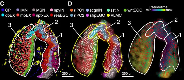
The introduction of stereo-seq to the study of axolotl brain regeneration has allowed scientists to examine the precise molecular events that occur during this process. By applying stereo-seq to axolotl brain tissue, researchers can map gene expression patterns in individual cells while simultaneously preserving the spatial context of the tissue. This enables them to see which genes are active in specific regions of the brain and, more importantly, which genes are involved in the regenerative processes that allow axolotls to regrow brain cells.
The breakthrough stereo-seq study on axolotls focused on several key aspects of brain regeneration, such as:
Cellular Identity and Differentiation: By capturing the gene expression of individual brain cells, researchers were able to identify distinct populations of cells involved in regeneration. This includes understanding the role of radial glial cells and how they differentiate into neurons during brain repair.
Molecular Pathways: Stereo-seq allowed scientists to pinpoint which molecular pathways are activated in response to brain injury. For example, certain genes related to inflammation, cell proliferation and cell differentiation were found to be upregulated in response to injury, suggesting they play a role in the brain’s regenerative process.
Spatial Mapping of Gene Expression: By mapping gene activity in precise spatial locations, researchers could see how different regions of the brain respond to injury. This is critical for understanding how axolotls can regenerate specific brain areas and how this knowledge can be applied to human medicine.
The Stereo-seq Approach, a New Era of Cellular Mapping:
The stereo-seq technique offers several advantages over previous methods used to study tissue regeneration.
HIGH SPATIAL RESOLUTION
One of the biggest challenges in studying tissue regeneration is capturing the precise location of gene activity within a complex tissue structure. Traditional methods, such as bulk RNA sequencing, provide valuable data on gene expression but lack spatial resolution. With stereo-seq, researchers can pinpoint the exact locations within the tissue where specific genes are active. This is particularly important when studying tissues like the brain, where cellular organization is critical to understanding function and regeneration.
INTEGRATION OF SPATIAL AND TRANSCRIPTOMIC DATA
Stereo-seq integrates both spatial and transcriptomic data, allowing researchers to simultaneously visualize the architecture of the tissue and understand the molecular mechanisms at play. This combination provides a much richer and more complete picture of the regeneration process. By studying axolotl brain tissue with this integrated approach, scientists can map out the spatial distribution of cellular activities during regeneration and link them to specific molecular events.
SINGLE-CELL RESOLUTION
Stereo-seq operates at a single-cell resolution, which is essential for understanding the heterogeneity of cells involved in regeneration. In the brain, different types of neurons, glial cells and precursor cells may exhibit different gene expression patterns depending on their developmental stage or regenerative status. Stereo-seq allows researchers to investigate these differences at the level of individual cells, revealing a detailed picture of how various cell types contribute to brain repair.
NEW INSIGHTS INTO AXOLOTL BRAIN REGENERATION
The stereo-seq findings from axolotls have already provided several exciting insights into the regenerative processes at play in their brains:
1. Radial Glial Cells as Key Players in NeurogenesisThe study confirmed that radial glial cells are central to brain cell regeneration in axolotls. These cells, which are typically found in the developing nervous system, retain their regenerative potential in adulthood. Stereo-seq data revealed that radial glial cells not only act as a source of new neurons but also help maintain the architecture of the regenerating tissue.
Interestingly, the study also found that radial glial cells in axolotls exhibit gene expression patterns that are very different from those of radial glial cells in mammals, suggesting that there may be distinct molecular mechanisms at play in the axolotl’s regenerative response.
2. Activation of Regenerative Pathways
Researchers identified several molecular pathways that are upregulated during brain regeneration in axolotls. Some of these pathways are involved in cell proliferation, differentiation and tissue remodeling. These pathways include the Notch, Wnt and Hedgehog signaling pathways, which are known to play crucial roles in development and regeneration in other species as well.
Stereo-seq revealed how these pathways interact in different regions of the brain and how they help guide the regeneration process. By identifying these pathways in axolotls, researchers may be able to design targeted therapies that could encourage neurogenesis in humans, potentially offering new treatments for neurodegenerative diseases.
3. The Importance of Inflammatory ResponseAnother interesting finding from the stereo-seq study was the role of inflammation in brain regeneration. While inflammation is often considered detrimental to tissue repair, the data revealed that a controlled inflammatory response is crucial for promoting neurogenesis in axolotls. This suggests that inflammation may have a dual role, both aiding in the repair of damaged tissue and helping to guide the regenerative process.
IMPLICATIONS FOR HUMAN MEDICINE
The insights gained from studying axolotl brain regeneration using stereo-seq have significant implications for human medicine, particularly in the fields of neurology and regenerative medicine. By understanding the genetic and molecular mechanisms behind axolotl brain repair, scientists can identify potential therapeutic targets for encouraging brain cell regeneration in humans.
IN CONCLUSION
The first-ever axolotl stereo-seq represents a major leap forward in our understanding of brain cell regeneration. By combining spatial mapping with gene expression data, this technique has provided unprecedented insights into the molecular and cellular processes that drive axolotl brain repair. The findings from this study not only deepen our understanding of regenerative biology but also offer exciting new possibilities for treating brain injuries and neurodegenerative diseases in humans. As research in this field continues, we may one day be able to harness the regenerative powers of axolotls to help heal human brains.
Contribute to the TBI Times